Read Shoulder MRI with a Rock-Solid Systematic Approach
No credit card required.
Is that tendinosis or a tear? Torn labrum or just an anatomic variant? What are those alphabet soup acronyms like ALPSA or HAGL? Shoulder MRI can feel overwhelming at first, and important findings are easy to miss.
That is why we teamed up with expert MSK radiologists Dr. Angela Atinga and Dr. Rakesh Mohankumar to build a course that actually makes this click efficiently. Through a comprehensive introductory video, Dr. Atinga walks you through a clear, step-by-step outside-in approach to shoulder anatomy, showing you exactly what normal looks like and what you need to look for.
Then, you apply that knowledge across 42 high-yield, fully scrollable cases that cover the core pathologies you need to see. You get a full-screen PACS experience, live feedback from our AI Attending, and annotated answers so you can skip the guesswork. You start. You finish. You walk onto your next shift knowing exactly what you are looking at.
Who This Course Is For
R3–R5 residents and MSK fellows. Anyone who wants a systematic approach to rotator cuff tears, labral pathology, and post-operative shoulder interpretation. Particularly valuable for trainees rotating through orthopaedic radiology.
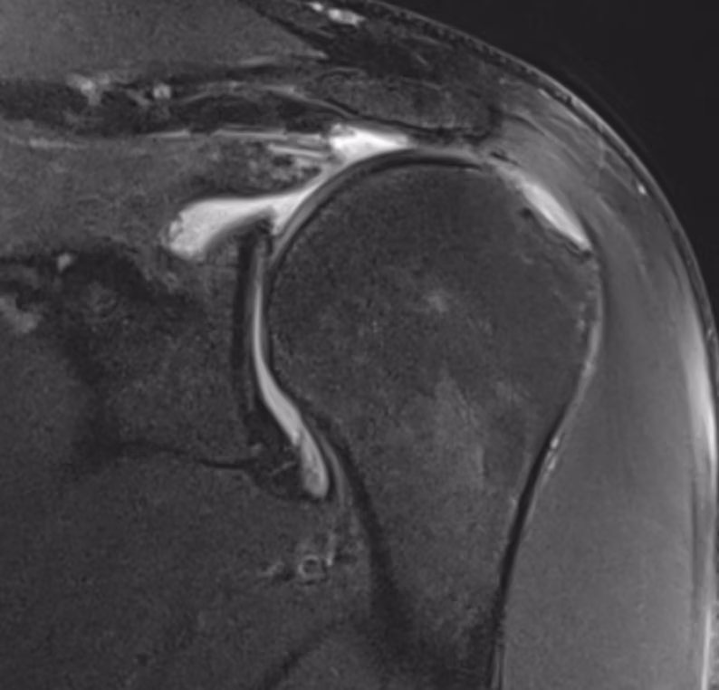
Pathologies include:
Get a taste for some of the pathologies covered in this course.
Normal Anatomy & Rotator Cuff
- ●Normal anatomy, arthrogram, and normal variants
- ●Partial thickness articular and bursal sided supraspinatus tears
- ●Full thickness supraspinatus tendon tear and massive rotator cuff tear
- ●Subscapularis tendon tear and delaminating intrasubstance infraspinatus tear
- ●Subacromial subdeltoid (SASD) bursitis and subcoracoid impingement
- ●Long head of biceps tendon dislocation and post-operative re-tears
Labrum and Instability
- ●Bankart and bony Bankart lesions
- ●ALPSA, Perthes, and GLAD lesions
- ●SLAP tears, posterior labral tears, and circumferential labral tears
- ●Hill Sachs, reverse Hill Sachs, and Bennett lesions
- ●HAGL (Humeral Avulsion of Glenohumeral Ligament)
Capsule, Ligaments, Muscle & Nerve
- ●Adhesive capsulitis (Frozen Shoulder) and internal/posterosuperior glenoid impingement
- ●Fatty atrophy of rotator cuff muscles secondary to massive tearing
- ●Suprascapular nerve entrapment (spinoglenoid notch cyst)
- ●Parsonage-Turner syndrome and quadrilateral space syndrome
Joint-Centred Disorders & Post-Op
- ●Glenohumeral joint osteoarthritis and focal chondral defects
- ●Synovial osteochondromatosis, PVNS, and septic arthritis
- ●Milwaukee shoulder and AC joint dislocation
- ●Anterior labral re-tear post-Bankart repair and post-Latarjet procedure evaluation
Other Challenging Cases
- ●Shoulder tumour (metastasis) from renal cell carcinoma
- ●Proximal humerus sarcoma with intra-articular extension
What You'll Learn
- 1Master a systematic, outside-in search pattern for evaluating the shoulder, starting with the rotator cuff and moving sequentially to the biceps tendon, labrum, capsule, and bones.
- 2Confidently differentiate between true glenoid labral tears and normal anatomical variants (like the Buford complex or sublabral foramen).
- 3Accurately classify partial and full-thickness rotator cuff tears while assessing for critical secondary signs like muscle fatty atrophy and specific nerve entrapment syndromes.
- 4Evaluate complex instability patterns, including Bankart, ALPSA, and HAGL lesions, to determine appropriate surgical management.
"It really simulates the real-world experience. It's not just sitting there studying — it's sitting there practising."
Frequently Asked Questions
Ready to start Shoulder MRI?
Continue Your Learning
Knee MRI
Read knee MRI with precision using 32 expert-curated cases covering menisci, ligaments, and bone.
View CourseSpine MRI
Master spine MRI with 22 curated cases covering degenerative disease, tumours, and cord pathology.
View CourseIn support of improving patient care, this activity has been planned and implemented by Navigating Radiology. Pinnacle Conference, LLC is jointly accredited by the Accreditation Council for Continuing Medical Education (ACCME), the Accreditation Council for Pharmacy Education (ACPE), and the American Nurses Credentialing Center (ANCC), to provide continuing education for the healthcare team.