Master CT Head & CTA Neuro for On-Call Readiness
No credit card required.
There's textbook learning, and then there's practical radiology. When you're alone in the reading room at 2 AM and the ER needs a read on a trauma or stroke, you don't have time to second-guess yourself. You need to know what you're looking at.
This course covers the essentials of on-call neuroimaging. Featuring 31 scrollable DICOM cases, we focus on the must-not-miss bread-and-butter emergencies. You start with an introductory video that breaks down foundational anatomy, vital windowing techniques to spot subtle blood, and a conceptual search pattern specifically designed to catch mass effect, herniation, bleeds, and strokes.
Every case is built to teach a principle. You commit to a read. The AI Attending guides your reasoning.
Who This Course Is For
R1–R3 residents preparing for neuro call shifts or a neuro rotation. Essential for anyone who has never confidently read a CT head independently — and for more senior residents who want to tighten their approach to subtle findings like early ischaemia or extra-axial collections.
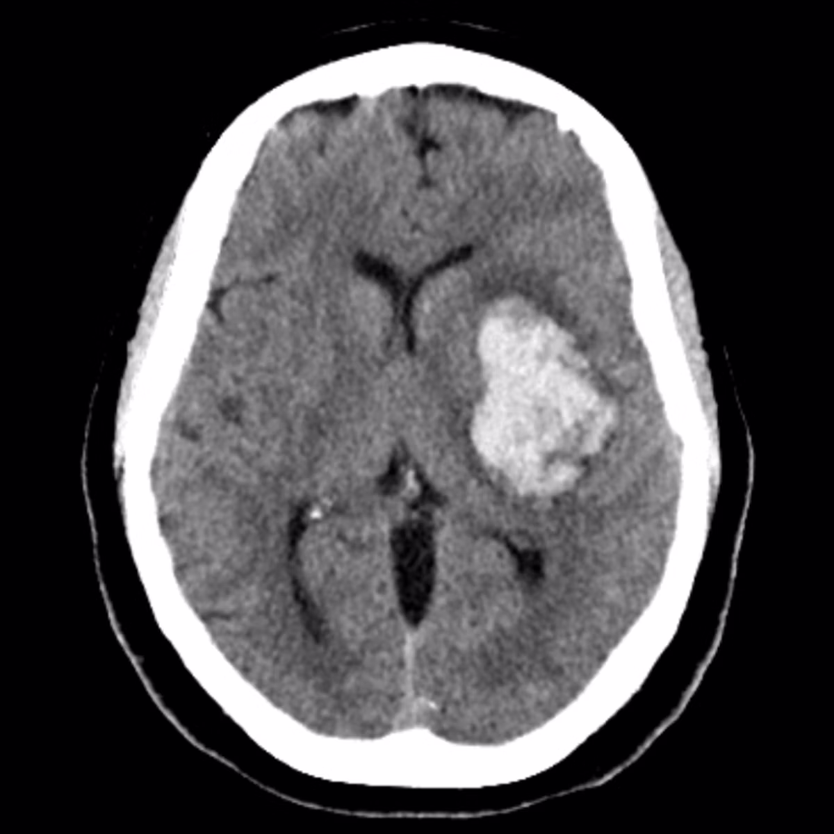
Pathologies include:
Get a taste for some of the pathologies covered in this course.
Stroke
- ●Including acute MCA infarct, vertebral dissection, and venous sinus thrombosis
Non-Traumatic Haemorrhage
- ●Including subarachnoid haemorrhage, hypertensive ICH, and aneurysmal rupture
Infection & Mass Lesions
- ●Including brain abscess, HSV encephalitis, and colloid cyst with hydrocephalus
CTA Neuro
- ●Including large vessel occlusion, Circle of Willis aneurysm detection, and vertebral artery dissection
What You'll Learn
- 1Systematically identify critical brain herniation syndromes (subfalcine, uncal, and tonsillar) and evaluate the basal cisterns for mass effect.
- 2Recognise the early signs of acute ischaemic stroke, including the hyperdense vessel sign and the subtle loss of grey-white differentiation.
- 3Properly manipulate CT windows (e.g. using a blood window) to detect subtle extra-axial haemorrhages that are easily missed on standard brain windows.
"I could have read five hours and gotten less than I got in 30 minutes. In terms of practical application, there's a gap between knowing things and being able to read a study. This bridges it."
Frequently Asked Questions
Ready to start CT Head & CTA Neuro?
Continue Your Learning
Brain MRI
Decode brain MRI with 67 expert-curated cases covering infarcts, tumours, inflammation, and more.
View CourseSpine MRI
Master spine MRI with 22 curated cases covering degenerative disease, tumours, and cord pathology.
View CourseCT Chest & CTPA
Navigate thoracic imaging with 40 real DICOM cases covering PE, dissection, diffuse lung disease, and more.
View CourseIn support of improving patient care, this activity has been planned and implemented by Navigating Radiology. Pinnacle Conference, LLC is jointly accredited by the Accreditation Council for Continuing Medical Education (ACCME), the Accreditation Council for Pharmacy Education (ACPE), and the American Nurses Credentialing Center (ANCC), to provide continuing education for the healthcare team.