Master CT Abdomen/Pelvis for On-Call Readiness
No credit card required.
Radiology is hard. You can read 100 pages about CT abdomen in a textbook, but when a real case comes through at 2 AM, the connection between what you read and what you see just isn't there. You shouldn't have to figure this out alone in the darkroom.
This course bridges that gap by teaching you from the ground up. We start with a foundational video covering essential anatomy and an approach, then apply that approach with 52 carefully curated, scrollable DICOM cases covering the bread-and-butter emergencies you should never miss — plus the clinical pearls that will take your reports to the next level.
You commit to findings. You submit your interpretation to the AI Attending. You get Socratic feedback. By the end, you won't just know what pathologies like appendicitis look like — you'll recognise them at 2 AM.
Who This Course Is For
R1–R3 residents preparing for body imaging call shifts, and physicians and surgeons who want to learn to read abdominal CT. Also valuable for international graduates who want to build their pattern recognition. Senior residents who want to tighten their systematic approach before fellowship.
Pathologies include:
Get a taste for some of the pathologies covered in this course.
Acute Hepatobiliary Pathologies
- ●Including acute cholecystitis, choledocholithiasis, hepatic abscess, and gallstone ileus
Acute Pancreatic Pathologies
- ●Including acute pancreatitis with necrotising complications and pseudoaneurysm
Acute Bowel Pathologies
- ●Including appendicitis, bowel obstruction, ischaemia, sigmoid volvulus, and intussusception
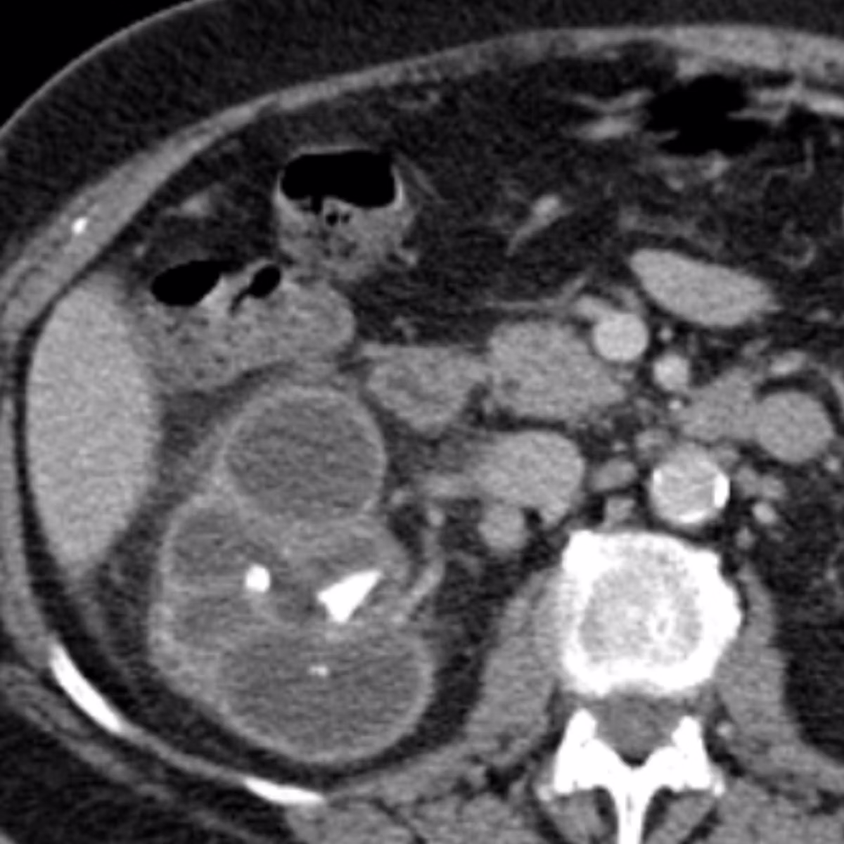
Acute Renal Pathologies
- ●Including pyelonephritis and obstructing ureteric stone
Acute Gynaecological Pathologies
- ●Including ovarian torsion
Lower Abdominal Pain
- ●Including Crohn's disease with active inflammation, epiploic appendagitis, and diverticulitis
What You'll Learn
- 1Systematically evaluate a CT abdomen/pelvis using a reliable, repeatable search pattern so you don't miss critical findings.
- 2Confidently identify complex peritoneal and retroperitoneal anatomy, including the spaces where free fluid and tumour metastases commonly hide.
- 3Recognise and report the imaging features of pathologies like appendicitis, pancreatitis, and bowel obstruction — and build the pattern recognition to identify them confidently in a PACS-style viewer.
"I saw a cystic artery pseudoaneurysm on a call shift. I recognised it immediately from the gallbladder module. My attending was surprised. Those kinds of moments have happened several times now."
Frequently Asked Questions
Ready to start CT Abdomen/Pelvis?
Continue Your Learning
CT Chest & CTPA
Navigate thoracic imaging with 40 real DICOM cases covering PE, dissection, diffuse lung disease, and more.
View CourseCT Post-Op Abdomen
Learn to read post-operative abdominal CTs systematically with 24 curated cases. It covers the principles and common complications you need to know.
View CourseIn support of improving patient care, this activity has been planned and implemented by Navigating Radiology. Pinnacle Conference, LLC is jointly accredited by the Accreditation Council for Continuing Medical Education (ACCME), the Accreditation Council for Pharmacy Education (ACPE), and the American Nurses Credentialing Center (ANCC), to provide continuing education for the healthcare team.