Navigate Biliary Pathology on MRI with Confidence
No credit card required.
Biliary pathology is everywhere in clinical practice. Obstruction. Cholangiocarcinoma. PSC. Gallbladder masses. Most trainees see these on CT but struggle when the hepatobiliary MRI comes through — because the protocol is more complex and the pathology overlaps.
This course cuts through that. Twenty carefully selected cases cover the five most important areas of biliary imaging: causes of obstruction, biliary tumours, gallbladder abnormalities, sclerosing cholangitis, and post-procedural assessment. Each case is built to teach a specific principle.
You commit to findings. You get immediate AI Attending feedback. By the end, you'll know how to approach the biliary tree systematically — and how to distinguish a benign stricture from a cholangiocarcinoma.
Who This Course Is For
R4–R5 residents. Body imaging fellows. Staff radiologists who want to be more confident with biliary MRI. Any radiologist who wants to stop guessing at biliary stricture characterisation.
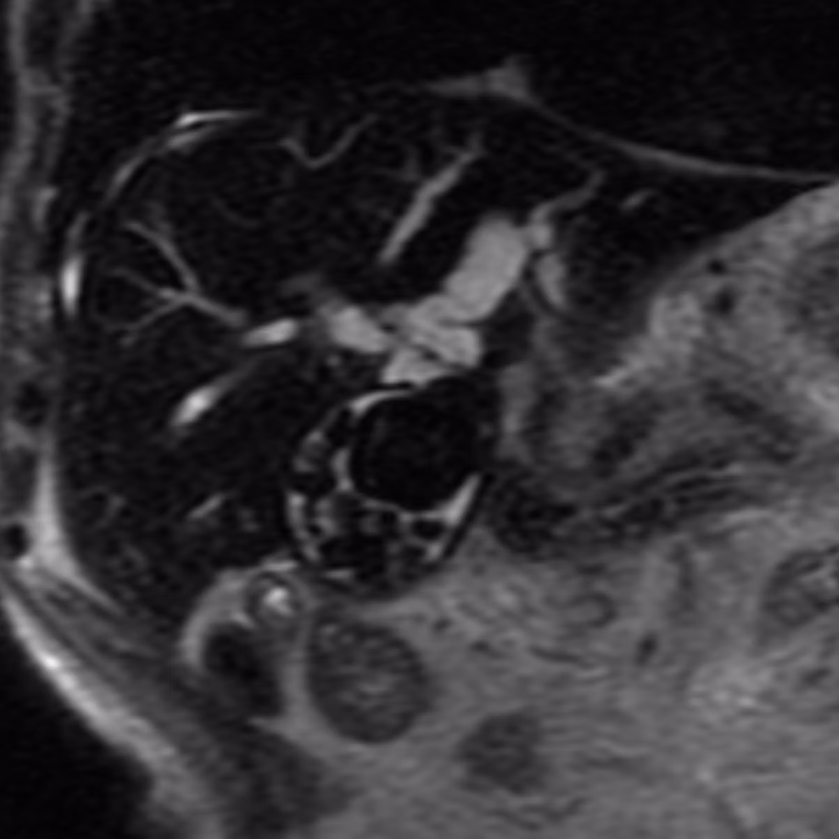
Pathologies include:
Get a taste for some of the pathologies covered in this course.
Biliary Obstruction
- ●Including choledocholithiasis, benign vs. malignant strictures, and Mirizzi syndrome
Biliary & Gallbladder Tumours
- ●Including hilar cholangiocarcinoma, distal cholangiocarcinoma, and gallbladder carcinoma
Sclerosing Cholangitis
- ●Including PSC and IgG4-related sclerosing cholangiopathy
Post-Procedure Complications
- ●Including bile leak, biloma, and anastomotic strictures
What You'll Learn
- 1Identify and classify the cause of biliary obstruction using MRI/MRCP — distinguishing stone disease, strictures, and neoplasm.
- 2Recognise and characterise cholangiocarcinoma in its three anatomical subtypes (hilar, distal, intrahepatic) and understand how location affects staging and surgical planning.
- 3Differentiate PSC from IgG4 cholangiopathy using specific imaging features and clinical context.
"It was like a teacher telling me: these are the important points, the rest you can forget. These are the things you should be looking at to keep a diagnosis or give a differential. It gave me direction."
Frequently Asked Questions
Ready to start Biliary MRI?
Continue Your Learning
Liver MRI
Master liver lesion characterisation with 30 real DICOM cases and expert video walkthroughs.
View CoursePancreas MRI
Master pancreatic mass characterisation with 20 real DICOM cases.
View CourseRenal MRI
Master renal mass characterisation with 25 cases covering Bosniak v2019 and solid renal masses.
View CourseIn support of improving patient care, this activity has been planned and implemented by Navigating Radiology. Pinnacle Conference, LLC is jointly accredited by the Accreditation Council for Continuing Medical Education (ACCME), the Accreditation Council for Pharmacy Education (ACPE), and the American Nurses Credentialing Center (ANCC), to provide continuing education for the healthcare team.