Characterise Adrenal Lesions with Confidence
No credit card required.
The adrenal incidentaloma is one of the most common findings in abdominal imaging — and one of the most mishandled. The question is almost always the same: adenoma or not? Chemical shift imaging, washout patterns, signal characteristics. There's a systematic approach that makes most cases straightforward.
This course, built with expert guidance from Dr. Satheesh Krishna, teaches you that approach. Eight carefully curated cases cover the core adrenal pathology you need to know: from the lipid-rich adenoma to the pheochromocytoma to the adrenocortical carcinoma. Small case count, high yield.
Who This Course Is For
R4–R5 residents. Body imaging fellows. Staff radiologists who want to be more confident with adrenal MRI. Any radiologist who wants to move beyond "probably an adenoma" to a systematic, evidence-based approach.
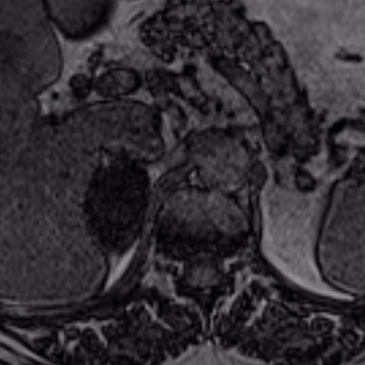
Pathologies include:
Get a taste for some of the pathologies covered in this course.
Adrenal Nodules & Adenomas
- ●Including lipid-rich adenoma, lipid-poor adenoma, and adenoma mimics
Benign Adrenal Masses
- ●Including myelolipoma, pheochromocytoma, and ganglioneuroma
Malignant Adrenal Masses
- ●Including adrenocortical carcinoma, adrenal metastasis, and adrenal lymphoma
What You'll Learn
- 1Apply a systematic approach to adrenal characterisation using chemical shift MRI — distinguishing lipid-rich from lipid-poor adenomas from non-adenomas.
- 2Recognise the key imaging features of pathologies like pheochromocytoma, myelolipoma, and adrenocortical carcinoma.
- 3Interpret adrenal lesions in the context of a known primary malignancy — distinguishing metastasis from incidental adenoma.
"Every money counts — coming from a different country, every payment seems like a lot. But it was the best investment I made. When I started my fellowship, I felt confident. I felt like I was part of the team."
Frequently Asked Questions
Ready to start Adrenal MRI?
Continue Your Learning
Renal MRI
Master renal mass characterisation with 25 cases covering Bosniak v2019 and solid renal masses.
View CourseLiver MRI
Master liver lesion characterisation with 30 real DICOM cases and expert video walkthroughs.
View CoursePancreas MRI
Master pancreatic mass characterisation with 20 real DICOM cases.
View CourseIn support of improving patient care, this activity has been planned and implemented by Navigating Radiology. Pinnacle Conference, LLC is jointly accredited by the Accreditation Council for Continuing Medical Education (ACCME), the Accreditation Council for Pharmacy Education (ACPE), and the American Nurses Credentialing Center (ANCC), to provide continuing education for the healthcare team.