CT Cervical Spine: An Approach for On-Call Confidence
No credit card required.
CT C-spine is one of the highest-stakes reads you will do as a trainee.
The combination of technical complexity, subtle fracture patterns, and the clinical consequences of a miss makes this a study where confidence matters. Most trainees know how to find an obvious fracture. But the subtle ones — the type II odontoid fracture, the Hangman's fracture, the occipital condyle injury — require a systematic approach that does not rely on luck.
This course is structured around a single comprehensive introductory video that covers everything you need to know: normal cervical spine anatomy, how to approach the study systematically, the key fracture patterns and their mechanisms, and high-yield cases worked through in video format.
DICOM cases for hands-on practice are in development and will be added to the course as they are completed.
Who This Course Is For
R1-R3 radiology residents on call who read CT trauma. Emergency medicine and trauma surgery trainees who want to understand C-spine CT interpretation. Any learner who wants a systematic framework before their first trauma call.
Pathologies include:
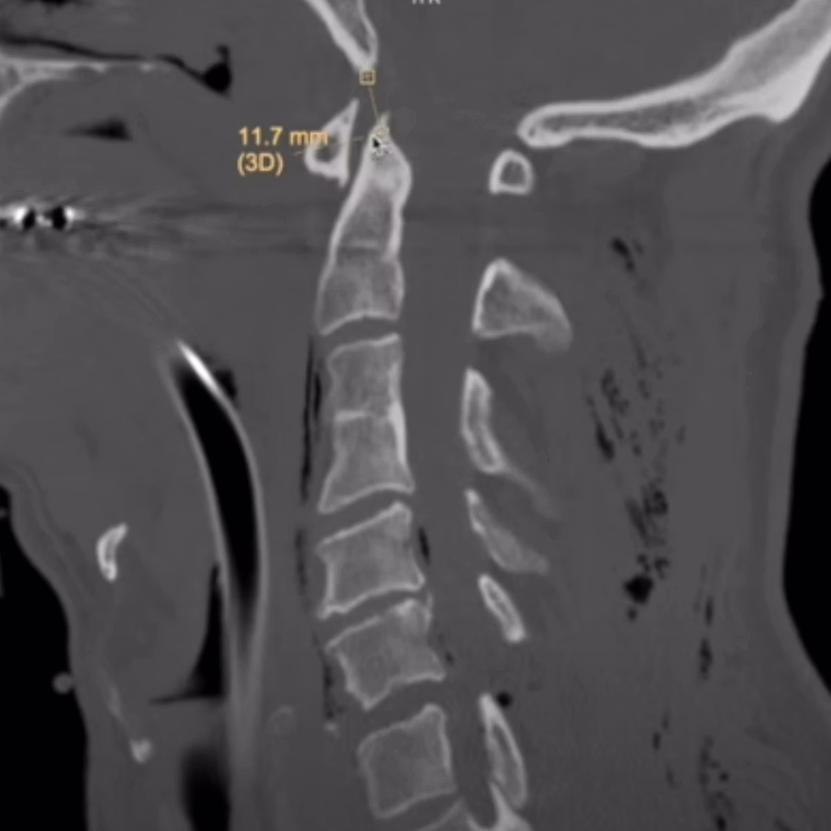
Get a taste for some of the pathologies covered in this course.
Key Fracture Patterns
- ●Including odontoid fractures, hangman's fracture, burst fractures, and teardrop fractures
Craniocervical Junction Injuries
- ●Including occipital condyle injuries and bilateral facet dislocation
Ligamentous & Soft Tissue Injury
- ●Including prevertebral soft tissue swelling and instability markers
What You'll Learn
- 1Apply a systematic approach to CT cervical spine that covers the craniocervical junction, vertebral bodies, facets, and posterior elements without missing subtle injuries.
- 2Recognise the key fracture patterns by mechanism — extension, flexion, axial load — so you can anticipate what you are looking for.
- 3Understand the clinical significance of each fracture type: which are stable, which are unstable, and which require urgent communication.
- 4Know when CT is not enough and when to recommend MRI for ligamentous injury assessment.
Frequently Asked Questions
Ready to start CT Cervical Spine?
In support of improving patient care, this activity has been planned and implemented by Navigating Radiology. Pinnacle Conference, LLC is jointly accredited by the Accreditation Council for Continuing Medical Education (ACCME), the Accreditation Council for Pharmacy Education (ACPE), and the American Nurses Credentialing Center (ANCC), to provide continuing education for the healthcare team.